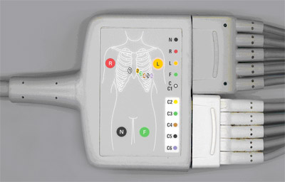
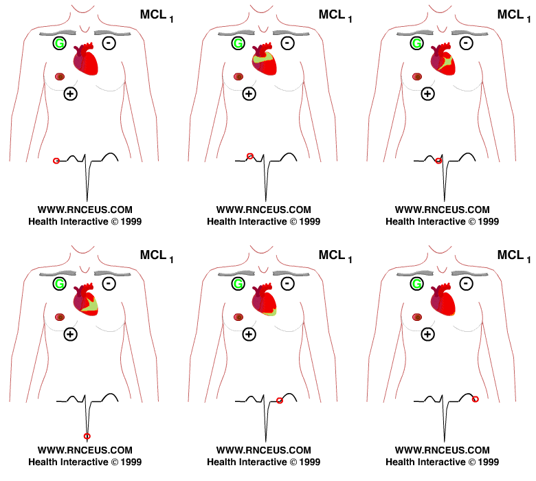
telemetry monitoring lead placement sorted by
relevance

Admin10.07.2021

Electrode Placement

Simultaneous processing of signals from 3 channels of ECG provides a multidimensional view of cardiac function, detecting changes that may not be visible in a single lead.
8608
Admin20.07.2021

ECG Lead positioning • LITFL • ECG Library Basics

The first time I really got my brain around getting deeper into 12-leads was when I was studying for my national critical care certification CCRN in 2015, five years into my career.
22010
Admin31.08.2021

Holter Monitor 5 Lead Placement Diagram

Rationale: The R wave should be approximately twice the height of the other components of the ECG to obtain proper detection by the heart rate counter in the equipment.
2400
Admin01.09.2021

How to use ECG

Detecting P waves in wide complex tachyarrhythmia to identify atrioventricular dissociation Lewis lead placement• They are called V1, V2, V3, V4, V5, V6, RA, LA, RL, and LL.
Admin02.07.2021

Holter Monitor 5 Lead Placement Diagram

Brady WJ, Lipinski MJ et al.
5602
Admin13.06.2021

5

The three-lead system is the oldest and simplest of all cardiac-monitoring lead systems.
8705
Admin29.08.2021

5

This is necessary because nurses on the floor are caring for many more patients and their patients are not on a bedside monitor for them to be able to visualize immediately.
807
Admin19.06.2021

Holter Monitor 5 Lead Placement Diagram

If the patient does not respond to IV fluids then further investigation is needed.
Admin01.08.2021

Elsevier

It takes some time to understand how to interpret 5-Lead ECGs, and specifically what you need to know for your unit.
4304
Admin20.07.2021

Electrode Placement

There you have it, 5 lead ECG placement simplified.
2809
Admin19.07.2021

5

You can also go through that list up at the top of normal sinus rhythm as well.
401